| Table of Contents | |
|
Case Report
| ||||||
| Primary malignant lymphoma of testis in a young immune competent adult: A case report | ||||||
| Vandana Rana1, Devika Gupta1, Rajat Jagani1, Giriraj Singh2 | ||||||
|
1Associate Professor, Armed Forces Medical College, Department of Pathology and Laboratory Science, Command Hospital, Pune, India.
2Associate Professor, Armed Forces Medical College, Department of Radio diagnosis and Imaging, Command Hospital, Pune, India. | ||||||
| ||||||
|
[HTML Abstract]
[PDF Full Text]
[Print This Article]
[Similar article in Pumed] [Similar article in Google Scholar] |
| How to cite this article |
| Rana V, Gupta D, Jagani R, Singh G. Primary malignant lymphoma of testis in a young immune competent adult: A case report. Edorium J Pathol 2014;1:1–5. |
|
Abstract
|
|
Introduction:
Primary testicular lymphoma (PTL) is a disease of elderly and is very rarely seen in young adults. In the era of human immunodeficiency virus (HIV) pandemic, changing age spectrum has been reported but it is an uncommon malignancy in immune competent young adult. Chances of misdiagnosis or delayed diagnosis are there due to same age group of presentation as that of germ cell tumors.
Case Report: A 35-year-old immune competent male presented with left testicular swelling for five months duration, he was being treated as a case of epididymo-orchitis. On fine-needle aspiration cytology impression of seminoma was favored but it turned out to be non-Hodgkin's lymphoma (NHL) of testis on histopathology following orchidectomy. There was no evidence of generalized lymphadenopathy or involvement of any other organ. On immunohistochemistry (IHC) studies diagnosis of diffuse large B cell lymphoma (DLBCL), non germinal cell type was made. Conclusion: Testicular lymphomas are rare and aggressive tumors and timely diagnosis in early stage helps the prognosis. Even though our patient was immune competent, in the era of human immunodeficiency virus (HIV) pandemic with changing age spectrum it is important to consider lymphomas in differential diagnosis of testicular tumors even in young patients. | |
|
Keywords:
Diffuse large B cell lymphoma (DLBCL), Human immunodeficiency virus (HIV), Primary testicular lymphoma (PTL), Immunohistochemistry (IHC), Lymphoma, Seminoma, non-Hodgkin's lymphoma (NHL), Testis
| |
|
Introduction
| ||||||
|
Primary malignant lymphoma of testis is rare and is predominantly a disease of elderly [1] . In the era of HIV infections, a higher incidence and broader age spectrum has been reported [2]. However, primary testicular lymphoma (PTL) in immune competent young adult is very rare. Chances of misdiagnosis or delayed diagnosis more so on cytology are there due to same age group of presentation as that of germ cell tumors. We report a case of PTL in a 35-year-old male who offered us initial diagnostic challenge on fine-needle aspiration cytology (FNAC). The patient underwent orchidectomy and based on histopathological and immunohistochemistry, diffuse large B cell lymphoma (DLBCL), of testis was diagnosed. | ||||||
|
Case Report
| ||||||
|
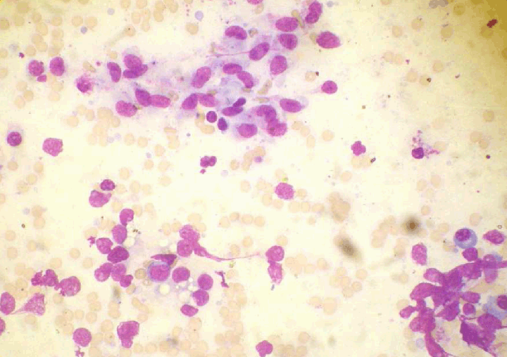
A 35-year-old male presented to our institute with complaint of enlarging left testicular mass for five months. There was no contributory history. Ultrasound done outside reported left testicular enlargement with hypoechoic and coarse echotexture and increased vascularity. Left hydrocele with thickened left epididymis was present. Impression given was of orchitis of infective etiology. He was treated as a case of epididymo-orchitis and was given trial of antitubercular treatment for one month outside. No abnormality detected on general and systemic examination. On local examination left scrotum was distended with enlarged testis measuring approximately 7x7 cm with normal overlying skin. Right testis was normal in size. Laboratory investigations revealed a normal hemogram, renal and liver function tests. LDH was raised to 530 IU/L. Tests for AFP and ß-hCG showed normal levels. Contrast-enhanced computed tomography (CECT) scan revealed heterogeneous enhancing soft tissue mass of 7x6.3 cm in left scrotal sac with thickened left epididymis and involved spermatic cord (Figure 1). Few left para aortic lymph nodes with largest measuring 1.5x1.2 cm visualized. All other solid organs were normal. Impression of testicular tumor of neoplastic etiology was given. FNAC done under guidance yielded limited blood mixed material and smears prepared showed mainly population of some transforming lymphoid cells including centroblasts, immunoblasts and mature lymphocytes along with few Sertoli cells and epithelioid like cells (Figure 2). No maturing stages of germ cells were seen. The possibility of seminoma was suggested. Decision for left high orchidectomy was taken in multispecialty tumor board. | ||||||
| ||||||
| ||||||
|
Pathology
| ||||||
|
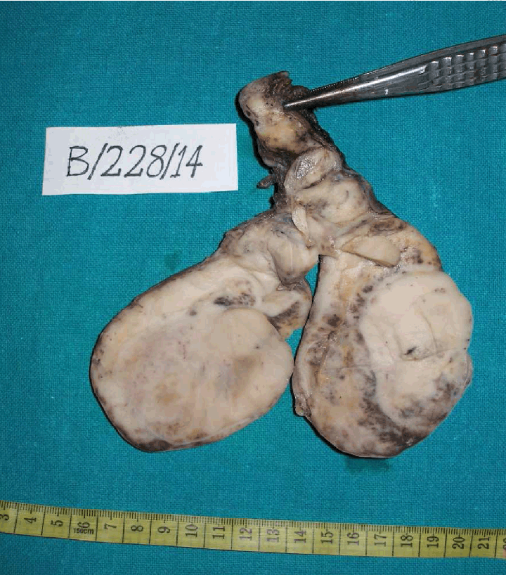
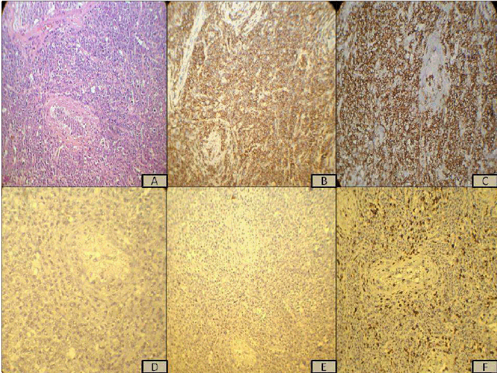
Received specimen of left high inguinal orchidectomy measuring 10x7.5x5 cm. Cut surface revealed a lobulated, grey-white fleshy tissue mass replacing most of the parent testicular parenchyma. Focal areas of hemorrhage seen but no necrosis was present. Epididymis and lower part of spermatic cord was grossly involved (Figure 2). On microscopic examination infiltrating population of large atypical lymphoid cells in sheets were seen replacing almost entire testicular parenchyma. Some residual hyalinized seminiferous tubules were seen encased by these atypical cells. The neoplastic cells were highly pleomorphic with high N:C ratio opened up chromatin and prominent nucleoli. Mitotic activity appeared brisk (Figure 3). Focal areas showed presence of mixed inflammatory infiltrate. The evidence of lymphovascular invasion was present. No evidence of intratubular germ cell neoplasm was seen. The tumor was seen infiltrating spermatic cord and forming nodules. Another separate specimen of an excised lymph node near deep inguinal ring was also submitted which was microscopically free of tumor and showed non-specific reactive changes. Immunohistochemistry panel of tumor cells revealed diffuse strong positivity for LCA, CD 20 and MUM 1 (Figure 4). Cells were negative for keratin, PLAP, CD117, CD 3, CD 5, CD 10 and BCL6. The diagnosis of DLBCL left testis, non-germinal center type was made. | ||||||
| ||||||
|
| ||||||
|
Discussion
| ||||||
|
Of all testicular tumors 94% are of germ cell origin and they occur primarily in young and middle aged adults [1]. The PTL is a rare disease and accounts for only 5% of all testicular neoplasm and represent 1–2% of all non-Hodgkin's lymphoma (NHL) [3]. It is the most common malignant tumor of the testis in elderly with average age being 60 years as proven both in old and recent studies [1, 4]. Few reports have shown increased incidence in younger age group more so in setting of HIV infections [2]. NHL is the second most common tumor in AIDS and is counted amongst the aids defining cancers. Incidence rates of NHL have been reported to be five times greater as compare to HIV pre epidemic period [5]. PTL in immune competent young male is very rare. Right and left sided testicular involvement is equal in frequency in PTL and approximately 6–38% of testicular lymphomas are bilateral [3]. Our patient is a 35 years old healthy male with unilateral testicular involvement and was in Ann Arbor stage IIAE. Young age, unilateral involvement and imaging studies pointed towards possibility of germ cell tumor in our case. On FNAC suggestion of seminoma was kept in spite of lack of tigroid background and characteristic large clear cells as seminoma is known to be associated with granulomatous reaction and rich lymphoid cell population [1]. Misdiagnosis is known, especially in cases of PTL with presentation in young age with seminoma and embryonal carcinoma being major mimickers [2]. Testicular lymphomas are more prone to misdiagnosis as germ cell tumors than vice versa [2]. IHC is essential in differentiating these tumors as treatment options vary in these entities. Histopathology and IHC studies lead to the diagnosis of PTL of DLBCL, non-germinal center type in our case. Curling first described a case of testicular lymphoma way back in 1866 [6]. Only approximately a decade later possibility of true PTL was considered keeping in mind the long-term survival in few patients following only orchidectomy [6]. PTL includes a heterogeneous group of lymphomas; DLBCL is the most common, comprising more than 70% of reported cases [2] [7] [8]. Other reported types include follicular lymphoma, plasmacytoma, lymphoblastic and Burkitt- like lymphoma [2] [7]. Most show B cell phenotype however cases of anaplastic T cell or natural killer cell lymphoma and rarely Hodgkin's disease has been reported [1] [2][7]. The majority of primary DLBCL of testis are of non-germinal center type that have high proliferative index [8]. Primary DLBCL of different immune privileged (IP) sites including testis have many common clinical and biological features like relatively poor prognosis, tendency to spread to other IP sites and deletion of HLA region [9]. PTL has aggressive clinical course and are known to involve extranodal sites at presentation and at relapse [7] PTL is reported to involve the skin and subcutaneous tissue in 6–13%, Waldeyer's ring in 4–6% and CNS in 3–6%. The central nervous system and Waldeyer's ring are common sites of involvement at relapse and carry a poor prognosis. Less common sites are the lung, bone, liver, GIT and nodal sites, especially the paraaortic lymph nodes [7]. Our patient is free of any distant metastasis. The outcomes in PTL have been poor, significant gains have been made with successive addition of radiotherapy, full course anthracycline based chemotherapy, Rituximab and central nervous system directed prophylaxis [10]. Statistical analysis has shown first line chemotherapy with Rituximab as a prognostic factor along with other factors like stage, LDH levels, ß2 microglobulin levels and international prognostic index (IPI) [10]. Our patient is treated with six cycles of R-CHOP, intrathecal methotrexate and RT to contra lateral testis. Patient is on follow-up at our institution and is disease free for last 12 months following therapy. | ||||||
|
Conclusion
| ||||||
|
Primary testicular lymphoma in young can lead to misdiagnosis or delayed diagnosis as presentation is in age similar to germ cell tumors. Immunohistochemistry confirmation is needed as treatment varies. Lymphoma should always be considered for differential diagnosis in testicular tumors even in young to avoid missing on an aggressive tumor. | ||||||
|
References
| ||||||
| ||||||
|
[HTML Abstract]
[PDF Full Text]
|
|
Author Contributions
Vandana Rana – Substantial contributions to conception and design, Acquisition of data, Analysis and interpretation of data, Drafting the article, Revising it critically for important intellectual content, Final approval of the version to be published Devika Gupta – Analysis and interpretation of data, Revising it critically for important intellectual content, Final approval of the version to be published Rajat Jagani – Analysis and interpretation of data, Revising it critically for important intellectual content, Final approval of the version to be published Giriraj Singh – Analysis and interpretation of data, Revising it critically for important intellectual content, Final approval of the version to be published |
|
Guarantor of submission
The corresponding author is the guarantor of submission. |
|
Source of support
None |
|
Conflict of interest
Authors declare no conflict of interest. |
|
Copyright
© 2014 Vandana Rana et al. This article is distributed under the terms of Creative Commons Attribution License which permits unrestricted use, distribution and reproduction in any medium provided the original author(s) and original publisher are properly credited. Please see the copyright policy on the journal website for more information. |
|
|
|
About The Authors
| |||
| |||
| |||
| |||
| |||